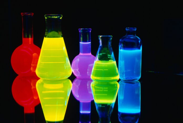
Chemiluminescence imaging is an innovative light-based technology that is revolutionizing how researchers visualize and analyze biochemical processes in living systems. This highly sensitive detection method is gaining popularity in biomedical research fields like molecular biology, immunology, and disease diagnostics.
What is Chemiluminescence Imaging?
Chemiluminescence is the emission of light from a chemical reaction. In chemiluminescence imaging, the chemical reaction produces a glow that can be detected and photographed with sensitive cameras. Researchers use enzymatic reactions that generate light to label molecules of interest in biological samples. The emitted light is then captured as an image on the camera, allowing visualization of the labeled target molecules or cells within the sample.
Several common chemiluminescent substrates generate light via oxidation reactions catalyzed by enzymes. For example, luciferase enzymes emit light during the oxidation of luciferin. Alkaline phosphatase enzymes produce light using chemiluminescent phosphate substrates. Horseradish peroxidase catalyzes light emission from enhanced chemiluminescence substrates containing luminol. The specificity of the enzymes ensures light is only produced where the target molecule is located, enabling sensitive detection and localization.
Advantages over Fluorescence Imaging
While fluorescence microscopy is the most widely used light-based imaging technique, chemiluminescence offers several advantages. Chemiluminescent reactions do not require an external light source for excitation, avoiding autofluorescence, photobleaching, and background signals generated by light exposure. This results in an exceptionally low limit of detection. Chemiluminescence also has a large linear dynamic range, allowing both weak and strong signal intensities to be accurately quantified within the same image. These attributes make chemiluminescence imaging highly sensitive and suitable for detection of molecules expressed at low levels.
Applications in Molecular Biology and Disease Research
Chemiluminescence Imaging has found numerous applications in biomedical research where high sensitivity detection is crucial. In molecular biology, it is commonly used to visualize nucleic acid expression patterns via in situ hybridization probes. Chemiluminescence-based affinity assays like Western blotting and ELISAs allow sensitive protein analysis. In immunology, it detects cell surface markers and intracellular signaling molecules via immunohistochemistry and flow cytometry. Chemiluminescence imaging also aids disease diagnosis – for example, detecting plasma biomarkers for conditions like cancer or infections.
Emerging Applications and Future Directions
Researchers are constantly exploring new applications and enhancements for chemiluminescence imaging. For instance, it enables sensitive imaging of biomolecules in tissue sections, enabling spatial localization within anatomical context. Dual-color chemiluminescence utilizing distinct enzyme-substrate systems permits multiplexing and co-localization studies. Advances in cameras now allow real-time chemiluminescence imaging of dynamic processes like intracellular calcium signaling in live cells. Combining chemiluminescence with microfluidic devices opens up potential applications in point-of-care diagnostics, personalized medicine, and environmental testing. Looking ahead, newer chemiluminescent probes and amplification strategies will likely further decrease detection limits, while coupling with advanced microscopy may grant super-resolution capabilities to this technique. Overall, chemiluminescence imaging shows great promise as a valuable tool for elucidating biological mechanisms and advancing disease research.
In summary, chemiluminescence imaging utilizes light-generating biochemical reactions to sensitively visualize molecular targets and cellular processes. Its inherent advantages over fluorescence make it well-suited for applications requiring high detection sensitivity, such as molecular analyses, immunohistochemistry, diagnostics and live cell imaging. With ongoing developments, this versatile imaging modality holds exciting opportunities to accelerate biomedical discoveries and improve human health.
Get more insights on:
Chemiluminescence imaging is a technique that uses chemiluminescent reactions to produce light, which is then captured using light-sensitive cameras or film. In chemiluminescence, two chemical reactants come together to form an intermediate excited state product that decomposes and emits a specific wavelength of light in the process. This emitted light can then be used for imaging applications.
The main advantage of chemiluminescence imaging over other imaging techniques like fluorescence is that it does not require an external excitation light source. The reactants themselves provide the energy needed to produce light emission. This allows for low background noise and high signal-to-noise ratios. Chemiluminescence reactions can also produce a wide range of wavelengths from visible to near-infrared, making them compatible with a variety of cameras and optical filters.
Mechanism of Chemiluminescence Reactions
Most commonly used chemiluminescence reactions involve the oxidation of an organic molecule in the presence of oxidizing agents like peroxides or oxalates. The organic molecules are called chemiluminescent probes or labels. Two of the most widely used classes of chemiluminescent probes are dioxetanes and acridinium esters.
In a dioxetane-based reaction, thermolysis or hydrolysis of the dioxetane ring leads to the formation of an excited carbonyl product. This product then decomposes and emits a photon as it relaxes back to the ground state. Acridinium ester chemiluminescence involves the oxidative cleavage of an endoperoxide moiety to form an excited acridinium ion. Emission occurs as this ion decays to the ground state.
Applications of Chemiluminescence Imaging
The ability to produce light from chemiluminescent reactions has enabled their use in a variety of imaging applications. Some major uses of chemiluminescence imaging are:
Bioluminescence Imaging
Bioluminescent proteins and genes have been introduced into cells and model organisms to study cell signaling, protein interactions and metabolic pathways in live animals. Firefly luciferase is a commonly used bioluminescent label.
Immunoassays and Blotting
Chemiluminescent probes conjugated to antibodies allow sensitive and quantitative detection of proteins, nucleic acids, hormones etc. in Western blots, ELISAs, lateral flow assays and microarrays. Applications range from disease diagnosis to food/water testing.
Microarray Imaging
DNA microarrays use chemiluminescent labels to detect thousands of genes simultaneously and analyze gene expression patterns. Protein microarrays also employ chemiluminescence for profiling antibody-antigen interactions.
Pathology
In situ hybridization and immunohistochemistry techniques use chemiluminescent probes for localized detection of DNA, RNA and proteins in tissue sections, enabling disease diagnostics and research.
Luminescent compounds have been developed for intraoperative tumor imaging applications as well.
Advantages of Chemiluminescence Imaging
The primary advantages of Chemiluminescence Imaging can be summarized as:
– Does not require an external excitation light source, allowing for low autofluorescence and high signal-to-noise imaging.
– Wide dynamic range exceeding that of radioactive, fluorescence or colorimetric methods. Signals can be detected over several orders of magnitude.
– Compatible with a variety of detection platforms like cameras, film and plates due to the availability of probes emitting different wavelengths.
– Highly sensitive – can detect attomolar levels of analytes. Suitable for applications requiring low detection limits.
– Provides stable signals over long durations, unlike bioluminescence which decays rapidly. Suitable for time-lapsed imaging.
– Safer alternative to radioisotopic methods with no special equipment or licensing needed.
Recent Advances and Future Prospects
Recent advances have focused on expanding the palette of chemiluminescent probes, improving their brightness and photostability. Caged chemiluminescent compounds activated by uncaging have been developed for spatially controlled imaging applications. Multicolor reporters have also been generated through rationally designed probes.
Use of novel delivery systems like nanoparticles, polymers and hydrogels could further enhance chemiluminescence signals and enable new in vivo and ex vivo applications. Expanding the mechanistic diversity of chemiluminescent reactions through biomimetic and abiological approaches may lead to probes with better performance.
Integration with microfluidics, 3D printing and other technologies could miniaturize chemiluminescence assays. Deeper understanding of biochemical luminescence in nature may inspire novel probe design as well. With continued innovation, chemiluminescence imaging seems poised to find wider adoption across life sciences research and clinical diagnostics.
Get more insights on: Chemiluminescence Imaging

